Eyelid ptosis
What is ptosis?
The word refers to the displacement ptosis or drooping of an organ or anatomical part. In this case we speak of ptosis or blepharoptosis. In ophthalmologic jargon, the term ptosis usually refers to the eyelid. It may be unilateral or bilateral and can affect any age.
Why are its causes?
The most common causes are due to age, in which there is a detachment of the muscle that raises the eyelid.
Less common causes are congenital, neurological and trauma causing a damaged levator muscle.
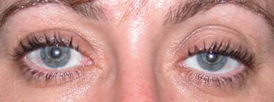
The use of contact lenses may predispose to the development of this condition.
 |
| Blepharoptosis involving the left eye in contact lens wearer |
What are the symptoms?
The main problem is that it can cause visual field loss, as more pronounced descents affect the visual axis.
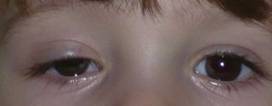
In children it can cause amblyopia, lazy eye, and sometimes needs to be dealt with at an early age.
 |
| Congenital blepharoptosis 3 year old with involvement of the visual axis OD. Requires treatment to prevent the amblyopic eye |
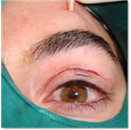
In adults there is a very important cosmetic reason, given that great asymmetry may occur.
 |
 |
| Ptosis in right eye lacking vision. Cosmetic purpose | |
How is ptosis treated?
The only treatment to correct ptosis is surgery.
There are several techniques described, which basically attempt to adjust the upper eyelid levator muscle to its original insertion.
Cases of severe functional impairment may need to be treated by implanting biological grafts, the patient, donor or artificial muscles anchored to rise against the eyelid. (Frontal suspension).
The goal of surgery is to leave open the visual axis, preventing loss of sight, leaving a palpebral margin that will fulfill its function of protecting the corneal surface. Therefore it must be harmonious, symmetrical and aesthetically acceptable.
The failure rate of recurrence or the causes may vary, being more common in congenital cases, children and those with a purely muscular etiology, in which it can be up to 20%.
The patient needs to be informed that the eyelid may return to its original position years after surgery.
 |
 |
| Blepharoptosis involving the left eye | |
| BEFORE | AFTER |
 |
 |
| Blepharoptosis involving both eyes. Reapplication using the superior levator muscle | |
| BEFORE | AFTER |
 |
 |
| Blepharoptosis involving both eyes | |
| BEFORE | AFTER |
Surgery is performed under local anesthesia and sedation. In children general anesthesia should be used.
 |
 |
| Congenital blepharoptosis involving the right eye | |
| BEFORE | AFTER |
 |

|
| Frontal suspension with fascia lata in a patient with congenital blepharoptosis without muscle function | |
Surgery usually lasts 30-60 minutes. It is not painful and does not require hospitalization.
Generally external sutures are hidden in the sulcus, leaving no scars.
In cases where the suspension is to be carried out to the front, the incisions are performed near or above the eyebrow and may leave a scar, which in any case will be small.
After surgery the patient should follow post-operative care advine as explained by the surgeon.


